
Weight, length, and head circumference are among the first measurements taken at birth and represent fundamental tools of neonatal auxology, the discipline that studies bodily growth and development. These are not simply numbers to be entered into the medical record: their interpretation makes it possible to place the newborn in relation to the reference population, evaluate their body proportions, and track their growth trajectory over time.
Neonatal growth, however, cannot be interpreted on the basis of a single value. What carries the greatest clinical significance is the trend over time, observed through correctly performed measurements and plotted on curves appropriate for sex, gestational age, and postnatal age.
The Italian Society of Neonatology (SIN), through the Perinatal Auxology Study Group, pays special attention to the anthropometric evaluation of the newborn. The INeS charts (ItalianNeonatal Study) represent one of the Italian reference standards for assessing newborn size at birth and were constructed as part of a prospective study involving over 45,000 newborns in 34 Italian centers. These charts take into consideration gestational age, sex, and birth order. The anthropometric measurement therefore constitutes the starting point of a surveillance journey that continues through the first months and years of life.

Weight: a dynamic measurement
The weight is probably the parameter most frequently used to track newborn growth, but it is also the one most influenced by physiological variations in the first days of life.
At birth, weight reflects numerous factors, including gestational age, genetic inheritance, fetal growth, maternal and placental conditions, and the course of the pregnancy. For this reason, the absolute value expressed in grams must always be interpreted in relation to gestational age and sex.
A newborn of 2,800 grams born at 39 weeks, for example, cannot be evaluated in the same way as a newborn of the same weight born at 32 weeks. It is precisely for this reason that neonatal curves report anthropometric parameters as a function of gestational age.
The INeS charts of SIN were developed for this purpose and can be used for newborns with a gestational age between 23 and 42 weeks.
Physiological weight loss
In the first days after birth, it is physiological to observe a reduction in body weight, a phenomenon mainly linked to fluid loss and changes in fluid balance that accompany the transition from intrauterine to extrauterine life.
The amount of weight lost and subsequent recovery must, however, be interpreted within the overall clinical context, considering feeding, diuresis, clinical conditions, and mode of delivery.
The truly informative data is therefore not just how much the newborn weighs on a given day, but how their weight evolves relative to the expected trajectory.
From physiological loss to the resumption of growth
After the initial adaptation phase, weight progressively increases again. In the healthy term newborn, the growth velocity is particularly high in the first months and subsequently tends to decrease.
The World Health Organization (WHO) has developed specific standards for weight, length, and head circumference and, separately, for the growth velocity of these parameters. These standards derive from the MulticentreGrowth Reference Study, which followed healthy children from different populations and made it possible to describe growth from birth to the first years of life. This aspect is particularly important: a growth curve does not merely take a snapshot of body size, but describes a trajectory.
Two children can have the same weight at a given age and subsequently follow different paths. One can maintain a relatively stable position with respect to the curve over time, while the other can cross several percentiles upwards or downwards. It is precisely this modification of the trajectory that warrants clinical attention.
Length: linear growth
Length represents an indicator of skeletal growth and must be interpreted together with weight and head circumference.
In newborns and infants, it is measured in the supine position, using a specific instrument called an infantometer. The procedure requires correct positioning of the head, extension of the legs, and contact of the feet with the measuring surface. An apparently simple measurement can therefore be influenced by the technique used. Small differences in the child’s position can modify the value obtained, especially when comparing successive measurements. For this reason, in pediatric auxology, it is important to use standardized procedures and appropriate instruments.
The INTERGROWTH-21st project, in which Italy also participated through the Turin center, adopted standardized anthropometric protocols for weight, length, and head circumference, with the goal of obtaining comparable international standards across different populations.
Weight and length must be interpreted together
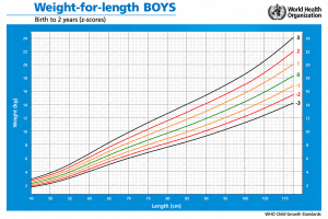
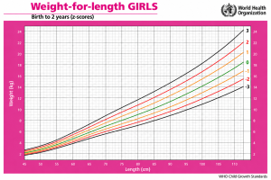
Weight, considered in isolation, does not fully describe the characteristics of growth. For example, a child with a high weight may also be particularly long and therefore present different body proportions compared to a child of the same weight, but of shorter length.
For this reason, auxological assessment includes multiple parameters and, when indicated, also ratios such as that between weight and length.
Head circumference: a measurement of brain development
The head circumference, often indicated by the acronym HC, is a particularly relevant measurement in the early period of life, as it indirectly reflects the growth of the skull and accompanies, at this stage, that of the brain. The measurement is taken by placing a non-stretchable measuring tape around the maximum circumference of the head, passing anteriorly through the supraorbital region and posteriorly through the occipital prominence.
In this case as well, standardization of the technique is crucial. An apparently minimal variation can in fact take on a different meaning depending on whether it represents a real change in growth or a difference in the measurement method.
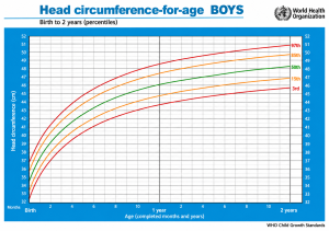
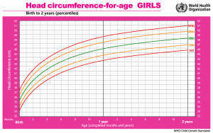
Head circumference is therefore interpreted in relation to age, sex, and the reference curve used. WHO standards include specific curves for this parameter and tools to assess its growth velocity.
Why head circumference should not be overlooked
In the first months of life, brain growth is particularly rapid. For this reason, head circumference is one of the parameters that the pediatrician considers during well-child visits. A single measurement, however, does not allow us to determine on its own whether a problem is present. It is the trajectory of head growth that provides more useful information. A value consistently located on a specific percentile can represent a normal individual characteristic, whereas a significant and persistent variation in trajectory requires a clinical evaluation.
Therefore, for head circumference as well, the trend over time takes on greater significance than the single value.
Percentiles: how to interpret them correctly
When weight, length, and head circumference are plotted on a growth chart, the child’s values can be compared with those of a reference population.
The percentile is not a grade and does not indicate, on its own, whether a child is healthy or has a pathological condition.
A child who falls on the 10th percentile for weight is not necessarily underweight, just as a child on the 90th percentile is not automatically overweight.
Interpretation must take into account several elements:
- Gestational age at birth and sex of the child.
- Birth weight and size, together with subsequent measurements.
- Growth trend over time, considering any trajectory variations.
- Ratio between weight, length, and head circumference, for an overall evaluation.
- Feeding and clinical conditions, which can influence growth.
- Family history, in relation to constitutional characteristics and growth patterns.
It is therefore more correct to consider the child’s position and trajectory on the curve, rather than attributing pathological significance to a single percentile.
Fetal growth and growth after birth are not the same thing
A particularly important aspect concerns the distinction between intrauterine growth and postnatal growth.
At birth, weight, length, and head circumference are compared with specific standards for gestational age. The INTERGROWTH-21st project has developed international standards for these parameters, stratified by gestational age and sex.
The standards were built in eight geographical areas and also included Turin, making the project particularly relevant for Italian neonatological practice.
After birth, however, the evaluation becomes longitudinal: it is no longer just the size reached by the child that matters, but also the speed at which they grow and the continuity of their trajectory.
Term infant and preterm infant: two different situations
In the preterm infant, auxological assessment requires particular attention. A baby born well before term has spent outside the womb part of the period during which, under physiological conditions, fetal growth would have continued. Furthermore, during neonatal hospitalization, illnesses, parenteral or enteral nutrition, infections, therapeutic interventions, and other conditions capable of modifying growth can intervene.
For these reasons, the assessment of preterm infant growth cannot simply be superimposed on that of term infants.
SIN draws attention to the use of appropriate curves for monitoring preterm infants and indicates, among the available tools, the INTERGROWTH-21st longitudinal standards for the postnatal growth of preterm infants. Furthermore, in follow-up, interpretation must take into account corrected age, especially in the first years of life.
Small, appropriate, or large for gestational age
The anthropometric measurements at birth allow classifying the newborn in relation to gestational age.
In general terms, three categories are distinguished:
- SGA – Small for Gestational Age: newborn small for gestational age.
- AGA – Appropriate for Gestational Age: newborn appropriate for gestational age.
- LGA – Large for Gestational Age: newborn large for gestational age.
This classification does not automatically equate to a diagnosis of disease.
A newborn can be constitutionally small or large without presenting a pathological condition. On the contrary, an alteration in growth can represent the expression of conditions that require further investigation.
SIN emphasizes precisely the importance of having an accurate gestational age and accurate measurements of weight, length, and head circumference at birth, so as to allow an appropriate auxological classification.
Growth is not linear
One of the most important concepts to convey to parents is that newborn growth does not proceed in a perfectly regular pattern. There are physiological variations in growth velocity and periods characterized by greater or lesser increases can occur.
The WHO describes a particularly high growth velocity after birth, followed by a progressive deceleration over the course of the first year of life. For this reason, it is not correct to expect the child to gain weight or length always at the same rate. Pediatric evaluation must therefore be based on repeated measurements over time, preferably carried out using comparable methods.
When the curve changes trajectory
An aspect that deserves particular attention is any persistent crossing of multiple percentile channels.
A change in trajectory can be physiological at certain moments of development, especially in the first few months, when the child tends to progressively stabilize towards their own growth channel. Conversely, a significant and persistent deviation may require further investigation.
Interpretation must always take into account the entire clinical picture. Possible causes of a growth slowdown may include feeding difficulties, insufficient nutritional intake, absorption problems, chronic diseases, or endocrine and metabolic conditions.
There is therefore no single value that allows, on its own, to make a diagnosis. It is the combination of measurements, their evolution, and the child’s clinical condition that guides the pediatric evaluation.
Conclusions
The growth of the newborn must be considered as a continuous process, which requires careful observation and an overall evaluation.
At birth, the measurement of anthropometric parameters allows defining the child’s position relative to gestational age. In the following months, repeated measurements make it possible to verify whether the trajectory remains consistent with their individual path.
The Italian Society of Neonatology, through its perinatal auxology activities, has over the years emphasized the need to use appropriate references for the different stages of neonatal growth.
The INeS charts represent an Italian reference for evaluation at birth, while INTERGROWTH-21st and WHO offer international tools for specific phases of the auxological pathway.
The choice of growth curve is therefore not a simple technical detail, but an integral part of clinical interpretation, essential for understanding the pattern of growth and accompanying the child through the different stages of development.
